Are you a #CuriousMind?
Embark on a journey of curiosity and exploration with ONI! Discover more about our innovations and pioneering advancements by visiting oni.bio/curiousminds
Share this article:
One of the characteristics of a multicellular organism is communication between their cells. Throughout evolution, organisms acquired several mechanisms to allow them to send messages between different cells, near and far. One of the famous examples of these mechanisms is hormones.
Another curious example is extracellular vesicles (EVs), a phenomenon that is only being discovered in recent years, with much still to uncover. EVs are bubble-like structures, where bits of the cell content, e.g., proteins, RNA, etc. are engulfed by cellular membranes. While it is still unclear how exactly their production and release are regulated, more and more evidence suggests that they are essential for proper cell-cell communication.
One of the examples for this can be found in the crosstalk between EVs and viral infections. Viruses frequently manipulate EVs on several different levels: some viruses introduce their own genetic material into EVs of infected cells, a process that can be used to either spread the infection to other cells or to modify their immune response against the virus. Equally, other viruses target the infected cell’s EV production machinery, which limits the cell’s ability to fight the virus off. Thanks to the similarity between the mechanisms behind the production of viruses and the mechanisms behind the production of EVs, some studies even go further and suggest that the evolutionary origin of EVs is in the biology of viral infections.
EVs also play roles in many other processes both under normal conditions and in disease. A multitude of examples for the roles of EVs can be found in cancer research. These range from manipulating anti-tumor activity of immune cells, through promoting metastasis, to modifying the microenvironment of tumors, and more. EVs are very prominent in current cancer research, and many groups are utilizing them to develop both diagnostic tools and experimental treatments. One of the key questions about EVs in cancer is which cells are producing them. It’s easy to see why this question is important when considering them as a diagnostic tool, however, this question is also important when developing treatments to target EV production, as different cells will respond differently.

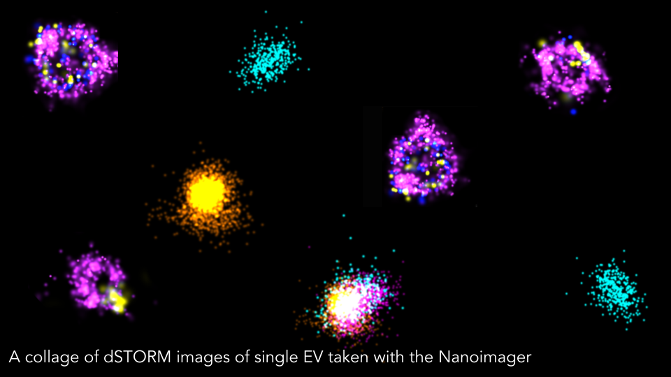
ONI recognizes the importance of EV in cutting edge research, and is therefore investing in developing tools to study EVs and to properly evaluate them. Our EV Profiler 2 kit is specifically designed to study EVs derived from human cells. After capturing and staining EV samples with our validated reagents, EVs are imaged in super-resolution using the Nanoimager, providing in-depth information about them. The combined qualitative and quantitative information provide invaluable insights into the nature of the EV sample. We are looking forward to seeing how our customers can take EV research to the next levels, and what strokes of brilliance we can still expect.
----------------------
Are you a #CuriousMind?
Embark on a journey of curiosity and exploration with ONI! Discover more about our innovations and pioneering advancements by visiting oni.bio/curiousminds
Share this article: