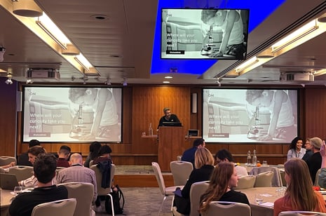
Scientists and researchers with an interest in microscopy and, especially, super-resolution imaging, came together in London for a second successful Super-Resolution Summit of the year - after the inaugural Summit which took place in June, in San Diego. During this occasion, the event gathered around 150 attendees, who had different levels of experience in super-resolution microscopy, and from a wide range of organizations, from both the public and private sectors.
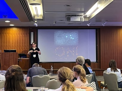
Amidst the vibrant atmosphere of knowledge exchange, networking, and thought-provoking discussions, the "Curious Minds" campaign from ONI was the glue bringing everything together during the Summit. Designed to ignite curiosity and foster innovation, the initiative aims to inspire researchers to push the boundaries of what is possible in scientific research and super-resolution imaging specifically. Through engaging sessions, interactive demos, and collaborative opportunities, the Super-Resolution Summit and the "Curious Minds" campaign together created an atmosphere of exciting biological exploration, propelling us into a future of limitless possibilities in the nanoscale world with the potential to have a great impact at all scales worldwide.
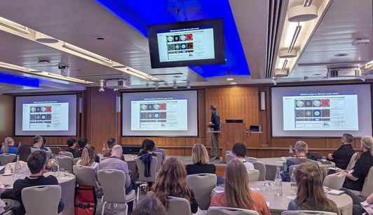
A highlight of the summit was the high-quality of the talks from thought leaders in this space, who shared their experience and latest results with the audience. Dr Lothar Schermelleh from the University of Oxford kicked off the sessions with a clear talk that covered the concepts of seeing, measuring and cross-validating is believing, beyond the classical “seeing is believing”. Lothar focused on the good all-around capabilities of Structured Illumination Microscopy (SIM), which can achieve sub-100 nm later and 250 nm Z resolution and has been used for different recent studies, including the study of single molecule Xist RNA dynamics or sororin monomer associated with cohesin (unpublished). He also highlighted the issue of oversharpening which can occur with the use of SIM2 or deconvolution.
On a similar line, Dr Susan Cox from King’s College London, who talked about finding ‘resolution’ within super-resolution, discussed some of the perils of artificial sharpening and making certain assumptions when using deconvolution or Fourier ring correlation analysis, one of the gold standards for measuring resolution. Susan enlightened the audience with her engaging explanations on different methods to decrease the likelihood of artificial sharpening, including the Haar Wavelet Kernel Analysis for simultaneous low and high density measurements, or their more recent work on the HAWK Method for the Assessment of Nanoscopy (HAWKMAN), a general approach that allows for the reliability of localization information to be assessed.
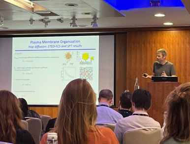
Following a productive networking break, the second session of the day had three very different and equally interesting talks centered around the importance of extracting meaningful data beyond single-molecule microscopy images. Firstly, Prof. Christian Eggeleing from Friedrich Schiller University Jena, shared their work on determining plasma membrane and molecular membrane dynamic heterogeneity. He covered his early work using Fluorescence Correlation Spectroscopy (FCS) and how they then combined it with STED, and separately used Single Particle Tracking (SPT) together with iSCAT to measure gold bead labeled lipids dynamics on the membrane, such as diffusion or compartment residence, among others. Christian presented their more recent work using SPT and MINFLUX, and a Python library called TRAIT2D to get over 10,000 tracks, with improved spatial and temporal resolution, and how they applied this to work with collaborators, such as Helge Ewers (HU Berlin), to obtain thousands of 3D tracks along neuronal axons of a GPI-anchored membrane protein. During the second half of his talk, Christian educated the audience on the concept of photoblueing, which means exposure to light can modify or photoconvert fluorophores resulting in a more blue emission, which they have seen can affect other dyes as well.
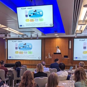
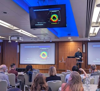
After that, Dr Daniel Rolfe from the Octopus Imaging Facility - STFC in Hartwell, inspired attendees with their use of single-molecule imaging and, in particular ONI’s Nanoimager, to assess EGFR oligomerization and create a fingerprinting tool with nanometer-resolution, which could impact the lives of many through precise drug design. The method that Dan and his colleagues have developed uses TIRF single-molecule localization microscopy and the FLImP method, whereby two photobleaching events are used to position two fluorophores close to each other, like in an EGFR dimer in this case, with around 10 nm resolution. So far, using 3 Nanoimager microscopes the team at STFC have managed to implement part of their vision to automate this workflow to help reveal new interfaces for drug treatment for non-small cell lung cancer - with ROI detection, thresholding, and autofocus refinement they have so far automatized data analysis, with >1.7 million cloud jobs and over 750 Tb data generated. Importantly, this method can be extended to MINFLUX and STORM-like data, also in 3D.
Concluding this session, Prof. Steven Lee from the University of Cambridge, explained how they use extra light to determine position, spectrum, and molecular orientation of each molecule. This ‘extra’ light comes from the nonlinear relationship between localization precision and photons detected, and using the end where extra photons do not bring much change to localization precision but can to determine other molecular parameters. He took us on a journey to understand how his team has used this concept to determine molecule orientation, a method coined POLCAM, which they have made available to the community through open-source image analysis software. For instance, POLCAM works in measuring the orientation of Nile red binding to alpha synuclein fibrils in vitro, or in dSTORM phalloidin AF647, for the polarized diffraction-limited imaging of membranes. Steven also discussed high-density volumetric super-resolution microscopy and its different approaches throughout recent history. He shared their work on Single Molecule Light Field (SMLFM), which exhibits superior resistance to high density datasets, and is eight-times faster than the Double Helix PSF current state of the art.
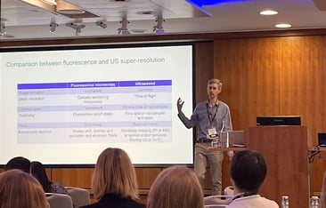
Last but not least, the last session of the day featured two scientists from different disciplines, showing how the super-resolution field benefits from a wide variety of expertises. First, Dr Vito Mennella from the University of Cambridge shared his work over the years in understanding the molecular architecture of centrosomes and cilia. Vito showed how complementary super-resolution techniques can be used to uncover molecular mechanisms and even new cellular structures that play crucial roles in airway tissue in both health and disease. For instance, their work on super-resolution microscopy and FIB-SEM imaging, which revealed parental centriole-derived, hybrid cilium in mammalian multiciliated cells, or their more recent unpublished work on ciliary rootlets architecture in beating cilia. To finalize a day of high-quality talks, Prof. Christopher Dunsby presented his work on ultrasound imaging to break the diffraction limit, as well as microbubbles (lipid shell with gaseous core) and nanodroplets (condensed microbubbles, 50-300 nm in size). They have created a model to localize single microbubbles, and then applied an improved system of two touching latex tubes imaged with a transducer to generate an ultrasound super-resolution image. Chris explained to the audience how, compared to fluorescence microscopy, ultrasound has coherent image formation, and depth resolved with time, with contrast from bubbles, flow or nanodroplet activation. Imaging in vivo using a mouse ear combined with microbubbles contrast agent, resolved vessels separated by 100 µm. The team can also measure bubble speed and flow direction. Finally, he highlighted how nanodroplets can rapidly be converted back into microbubbles via acoustics, which requires no flow, and their most recent work demonstrating Acoustic Wave Sparsely-Activated Localization Microscopy (AWSALM) and fast-AWSALM for in vivo super-resolution ultrasound imaging, which offers contrast on demand and vascular selectivity.
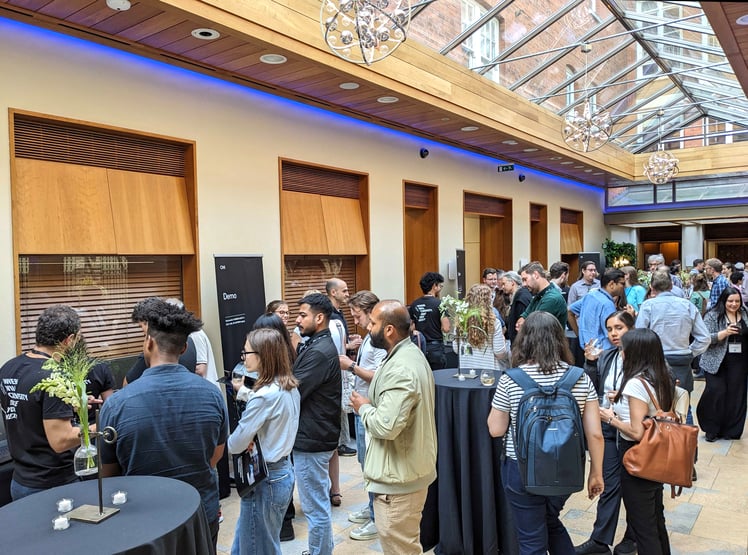
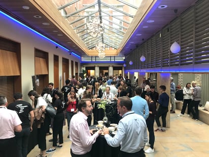
Besides the incredible talks, attendees had the opportunity to network with peers in the field working across a range of disciplines and applications, during both the breaks and happy hour sessions. For those curious about the Nanoimager and ONI’s product range, several members of ONI’s technical team were on site demonstrating with live samples, the Nanoimager’s capabilities to image cellular structures like mitochondria or nuclear pores with nanoscale-precision. The team also showed how ONI’s CODI software can help researchers extract crucial quantitative data from various datasets, including extracellular vesicles (EVs).
"The super-resolution summit truly embodies the essence of ONI. It seamlessly blends scientific discourse, captivating microscopy discussions, and an element of fun all in one event” said Carolyn Hartmann, Director of Product Marketing at ONI.
Altogether, the Super-Resolution Summit left a profound impact on all who attended. We are extremely thankful to everyone that took part and contributed to its success, including attendees, speakers, and organizers.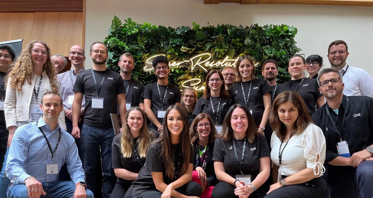
The journey doesn't end here! We invite you to join us at the upcoming summits in Boston on October 19, and Frankfurt on November 9, 2023. These events are your chance to immerse yourself in the forefront of super-resolution imaging, connect with peers and industry leaders in your area, and be part of groundbreaking technology. Don't miss out on this unparalleled opportunity to expand your knowledge and push the boundaries of what is possible in microscopy! Click here to learn more and secure your spot. We look forward to welcoming you as we continue this journey together!
Are you a #CuriousMind?
Embark on a journey of curiosity and exploration with ONI! Discover more about our innovations and pioneering advancements by visiting oni.bio/curiousminds