Characterizing Extracellular Vesicles (EVs) using conventional light microscopy can be quite challenging largely due to their small size, from 30 - 1000 nm in diameter. The challenge that we set when applying for the Nanoimager EV competition, was to characterize EVs captured inside a microfluidic device using dSTORM imaging.
In our lab we developed a microfluidics device, the Herringbone (HB)-chip, which can selectively isolate vesicles from a complex sample, using immunocapture of EV markers onto the chip’s surface. The EVHB-chip has complex physical properties that can be challenging when it comes to characterizing captured particles using super-resolution microscopy, such as the fabrication materials used and the 3D environment of the channels inside the device. We knew our project had many characteristics that would push the Nanoimager to its limits, and throughout the time we worked with close contact with the ONI technical support team so that we could ‘bend’ the system to achieve our goals.
Pushing the Nanoimager to its limits
In our lab we developed a microfluidics device, the Herringbone (HB)-chip, which can selectively isolate vesicles from a complex sample, using immunocapture of EV markers onto the chip’s surface. The EVHB-chip has complex physical properties that can be challenging when it comes to characterizing captured particles using super-resolution microscopy, such as the fabrication materials used and the 3D environment of the channels inside the device. We knew our project had many characteristics that would push the Nanoimager to its limits, and throughout the time we worked with close contact with the ONI technical support team so that we could ‘bend’ the system to achieve our goals.
The first challenge to overcome was the EVHB-chip material. The chip is made of COC plastic, with a bottom part being made of topaz polymer, which has a refraction index that is suitable for imaging. The Nanoimager works optimally with #1.5h glass coverslips, which are slightly different from our device. Nonetheless, even with this limitation we were able to image the herringbone structure inside the chip with some adjustments provided by ONI’s technical support team, such as alignment of the system lasers and adaptation of imaging parameters.
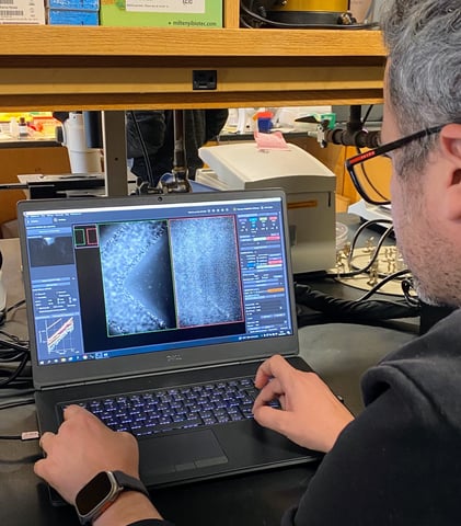
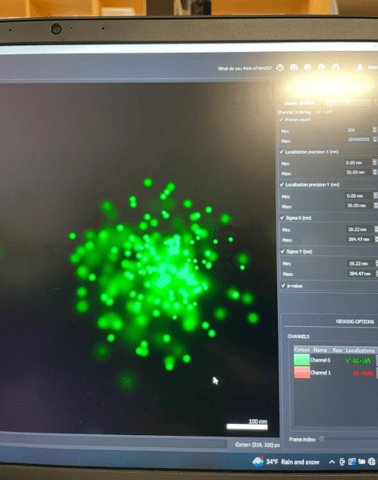
Figure 1. Image of the Stott’s lab custom made microfluidics HB-chip, taken with the Nanoimager and showing the herringbone pattern (left), and SARS-COV-2 particle captured on HB-chip (right).
More importantly, we were able to not just image on the first inner surface of the chip (equivalent to coverslip thickness), but up to 50 nm inside of the device channels. This is where our EVHB-chip has the highest capture efficiency, due to the presence of microfluidic patterns in the design that allow for enhanced mixing of the sample. It was fantastic to see that we could use the Nanoimager outside its default imaging definitions to achieve this. Despite all the incredible support from the ONI team, the specific characteristics of the chip still posed additional challenges to our imaging efforts. We were interested in imaging both SARS-COV-2 viral particles as well as EVs captured in our device, however, due to the limited time we were able to have the Nanoimager in the lab, we were unable to fully optimize our labeling and imaging process.
Nonetheless, we were able to image a SARS-COV-2 viral particle captured using one of ONI’s chips provided within the EV Profiler Kit. We functionalized the chip with the same antibodies that we routinely use in our lab to capture virus particles on the HB-chip surface, and labeled it with in-house conjugated antibodies. This showed the versatility of the ONI chips and their customization capability. One of the images generated have been included in a manuscript from the lab, which we expect to publish in the next few months.
Great products and support from the ONI team
Using the ONI EV Profiler Kit chips and provided reagents, we were able to access the expression of three tetraspanins (CD9, CD63, CD81) on the surface of EVs isolated in the lab using size-exclusion chromatography (SEC) columns.
We also found the CODI analysis software to be great for downstream data analysis and very user-friendly. We had previously used other super-resolution systems that did not provide such a simple experiment-to-result approach. Once again, wanting to be a bit less conventional, we used the ONI chips to look into characterizing rare markers on the EV surface. We used the capture antibodies provided by ONI and customized our labeling for these rare markers. Due to the lack of abundance of the marker of interest, we struggled with detection and analysis. Once again, the ONI support team was incredibly generous with their time and helped us throughout the whole process. Unfortunately, due to the characteristics of the sample we were unable to find a satisfactory number of clusters that would allow for analysis using the CODI software.
Overall, we were impressed with how user-friendly the software and the Nanoimager system are to use. Considering that it is a super-resolution imaging microscope, we were able to use it easily without needing much training or expertise. We would be incredibly grateful for all the support that we received from the ONI team, especially Ricardo Bastos, Alison Fujii and Raphael Jorand, who provided incredible on-site training and fantastic remote assistance throughout the 3 months’ period.
We would like to encourage other scientists in need of super resolution microscopy to apply for this year’s competition and get a chance to experience ONI’s platform first hand and for free for 3 months!
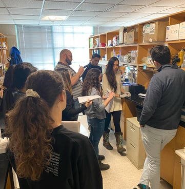
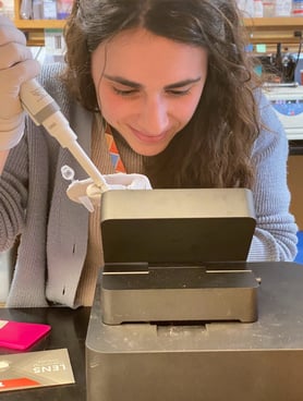
Figure 2. Members of the Stott lab enjoying their hands-on training session ahead of their 3-month period using the Nanoimager. Pictured are: Evelyn Luciano, Dasol Lee, Sara Cavallaro, Sara Veiga, Daniel Rabe, Raheel Ahmad, Daniel Ruiz and Alison Fuji.
------
Single-molecule localization microscopy, such as dSTORM imaging, on the Nanoimager enables the nanoscale resolution and precise localization of individual EVs, and their interactions with recipient cells. Further automated clustering analysis using ONI’s CODI platform could also be used to provide further insights and quantify EV populations, EV size, biomarker positivity and marker colocalization.
If you are keen to learn more about Nanoimager and how the ONI products can help your research, visit our EV hub or get in touch with us at hi@team.oni.