The International Society for Extracellular Vesicles (ISEV) annual conference took place in Kyoto from 24-28th April 2019. On behalf of the ONI team that had the pleasure to attend, I summarize here my impressions from the meeting, how the extracellular vesicles (EV) data generated at ONI was received and the latest developments taking place in the EV field.
Hello Japan!
On any other Monday, I would be going through my to-do list to prepare for the day at ONI’s Palo Alto office. However, at 11:56am on the 22nd of April, instead of sipping my morning coffee, I was boarding a bullet train at Tokyo station to reach the iconic city of Kyoto, and attend the annual International Society for Extracellular Vesicles (ISEV). I was excited to discuss our latest single-molecule vesicle data with other attendees, as well as meeting many of the people I had been talking to in the last few months.
-1.jpg)
Ready, set, go
Once settled in Kyoto, I prepared for Education Day, which reviewed major topics in EV research. The ISEV conference is the leading event for scientists in industry or academia working on anything related to extracellular vesicles (EVs). This year, almost 1200 attendees gathered for the annual event in the Miyakomesse convention center. The Education Day was incredibly well attended, with inspiring talks from the leaders in the field describing progress so far. Of particular interest were the MISEV guidelines on publishing EV studies, an exhaustive handbook on reporting experiments with the aim to improve reproducibility and standardization. Another highlight was the announcement of a second MOOC (Massive Open Online Course) for beginners in the EV field; the goal of the course is to provide basic knowledge about EVs. Here’s the link to the first MOOC, while MOOC 2 is announced!
-1.jpg)
Bottom picture credit: Dr. Kenneth Witwer, John Hopkins Medicine (shown during ISEV2019)
"Wait is that a vesicle? I thought it was a cell!”
The excitement was palpable throughout the conference, there was definitely a lot going on: plenary sessions, poster presentations, technical discussions – all happening at once, often making it hard to choose which sessions to attend. From an ONI team point of view, the response was overwhelmingly positive: at times, we felt our booth was really busy with people who wanted to know more about imaging single vesicles. One visitor, when shown an image of a single vesicle with membrane, CD63 and CD81 labeled, could not believe what he was seeing. He was in awe: "Wait, is that a vesicle?!", he thought it was a cell! (note to self: enlarge scale bar). People gathered around our screens to watch tracking videos of cell-vesicle interactions. Our presentation, given by Mariya Georgieva, also sparked considerable interest, with approximately 70 attendees. Numerous people came to ask us about the EV competition too – who would not want to use the Nanoimager, our unique super-resolution compact microscope, in their lab for two months? And for free?!
-Sep-23-2022-02-36-42-49-PM.jpg)
Moving forward
Besides the high-quality scientific contributions, the highlight of ISEV2019 for me, was the sense of curiosity, community, and collaboration that filled the halls at Miyakomesse. Everyone was eager to learn, share feedback and practices, while not being afraid to ask questions. A constructive atmosphere is indispensable in an emerging yet promising field, working together to accelerate clinical translation and meeting clinical needs. At the end of the meeting, the ISEV flag was passed on to ISEV2020 co-chairs Professors Languino and Weaver. I very much look forward to next year’s meeting in Philadelphia (and Lyon the year after!) to witness and contribute to the strides the extracellular vesicle field is taking.
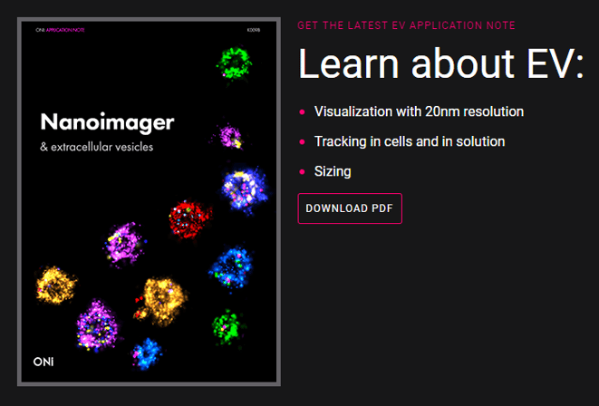
Download our application note on super-resolution imaging of extracellular vesicles using the Nanoimager.