FOM is a conference that gathers all scientists and companies working on the latest innovation and development in optical microscopy and their impact and application in material science, biology and medicine. The conference was a treasure of informative sessions covering a range of exciting topics such as single-molecule localization microscopy (SMLM), light-sheet, biomedical imaging, correlative microscopy, MINFLUX, and biophysics. It would be challenging to describe each single talk, with 5 exciting parallel sessions and more than 30 booths but below are some key talks to remark.
The conference began with tutorial sessions, among them the one led by Prof. Sjoerd Stalling, who provided a comprehensive overview of optics, super-resolution techniques and point spread function models. Many sessions covered light-sheet microscopy for large field-of-view and high-throughput acquisition. A particularly fascinating talk was the one introducing the Flamingo, a light-sheet microscope from Dr. Jan Huisken, who shared his vision of modular microscopes that are easy to rebuild according to the application, and easy to control. Dr. Isotta Cainero, from Prof. Diaspro’s lab, shared her work studying the organization of chromatin in neuro-degenerative disease using super-resolution techniques, such as Structured Illumination Microscopy (SIM) and Stimulated Emission Depletion microscopy (STED).On the third day, Prof. Elizabeth Hillman presented numerous applications based on their technology called SCAPE, a multispectral, high-speed, volumetric technology capable of imaging large-scale samples, such as rainbow worms or cleared and expanded mouse brains. The lab is now moving into medical applications by introducing the mediSCAPE design to replace conventional biopsies and histology with real-time imaging inside the living body.
Another microscopy topic presented in the conference was based on infrared and label-free technology, a technique that uses the auto-fluorescence of biological samples and avoids introducing fluorescence labeling, while penetrating deeper into the sample. Among those, Prof. Eric Potma shared his innovative work using non-linear optical microscopy in mid-infrared to determine cellular molecular composition, while Prof. Irene Georgakoudi showed two-photon metabolic imaging for the detection of cervical pre-cancers, with the exciting aim to translate into label-free clinical imaging.
On the last day, the conference was closed by two great plenary sessions of Prof. Melike Lakadamyali and Prof. Markus Sauer. Lakadamyali presented her latest work on a new machine learning model for segmenting and clustering point-cloud data to image tau protein in human postmortem brain tissues. Sauer exposed current localization-based microscopy limitations and introduced the 10 nm resolution barrier. He demonstrated that when fluorescent molecules are close to each other (less than 10 nm apart), the resonant transfer of energy leads to complicated photophysics, which induces lower localization probabilities that impedes fluorescence imaging at less than 10 nm… inviting users to be mindful about this!
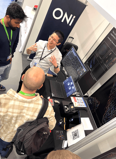
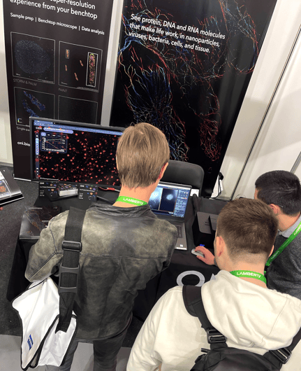
In parallel, the exhibitor space was also very busy, with plenty of companies showcasing their latest products and cutting-edge technologies. ONI had a booth, which drew many visitors. They were captivated by the Nanoimager's compact size and ability to perform real-time super-resolution imaging. Many were amazed by the on-site demonstration of nucleopore reconstruction, which added to the excitement and buzz around the booth! Many attendees also loved the walkthrough video of CODI displayed on the screen: which showed some of our tools in action, such as drift correction or clustering analysis, on the nucleopores’ sample.
“This is a very nice setup since users can use it themselves, without much maintenance. The data analysis platform is also great. We usually invest big once we choose a system”, said one of the visitors.
We had a great time at FOM and we look forward to joining the meeting and the microscopy community again next year. FOM 2024 will take place in Genoa - save the date on your diaries!
If you want to learn more about our complete solution for super-resolution imaging, visit our website or reach out to the team at marketing@oni.bio.