
The Science Innovation Union (SIU) at the University of Oxford hosted their first ever Art in Science Competition recently, with the aim to connect diverse scientific communities and provide scientists the chance of showcasing their research in the form of artwork. Sponsors, ONI, were at the award ceremony that took place on 10th May at the Department of Earth Science, University of Oxford.
75 submissions across different fields
Those scientists from within the SIU network wishing to display the art in their science in the form of scientific images, photographs and digital art sent their entries for the Art in Science competition. 75 submissions were received, which ranged from microscopy images, diagrams, graphs, protein models and other art-meets-science creations. It wasn't an easy task for the judges to pick the winner, although some stood out for different reasons.
The award ceremony
The event started with a brief presentation by the organizers, followed short presentations from the authors of the short-listed entries. ONI's Senior Applications Scientist Dr Ricardo Bastos gave a 15-minute presentation on ONI, our desktop super-resolution microscope, the Nanoimager, and the different imaging possibilities and applications it offers.
-1.jpg)
-1.jpg)
And the winner is...
Those attending the event were highly encouraged to vote for the People's Choice awards using an app, which represented two additional awards given out during the evening. Then, the winners of each category was announced; prizes were awarded in 6 different categories:
-1.jpg)
-
SIU Art in Science Winner: for Malte Kaller, PhD student at Nuffield Department of Clinical Neurosciences from the University of Oxford.
The image entitled "A failed experiment with aesthetic value" showed a human sensory neuron (in red), which was grown in a dish from re-programmed skin cells, with myelinating glia (in green); an interaction which is essential for the health and functioning of the nervous system. The nuclei were visualized in blue. The image was captured in a confocal microscope. Scale of the image is 320 x 320µm. The experiment was deemed unsuccessful, as the cell culture system was not optimal for the scientific question that he was trying to address, Malte's image caught the judges attention.
-
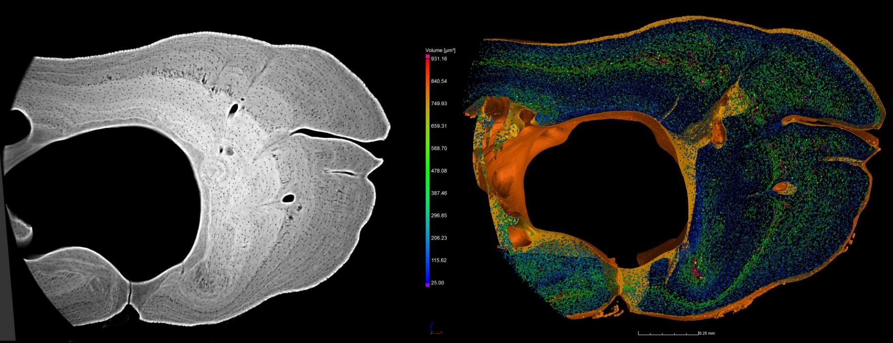
Best visual impact: for a joint submission from Armin Schmitt, Professor Roger Benson, Dr Donald Davesne and Marie-Christine Zdora (presented) from the Department of Earth Sciences at the University of Oxford.
Their mage "Bone Cells in the Rib of a Carp" shows a cross-section through the rib of a carp (Cyrinus carpio) (left), revealing a myriad of bone cells (osteocytes) inside the rib, seen as black dots. The annual growth cycles are visible as different shades of grey. The right part of the image shows a virtual 3D model of the osteocytes and the surface of the same bone. The different colors of the bone cells represent different size clusters; indicating a different volume of the bone cells during different growth stages of the animal. The scan was obtained using a sub-micrometer resolution at the ESRF light source in Grenoble, France.
The 3D rendering of the osteocytes was done with the VG Studio Max software. The scale bar on the image is 0.25mm.
-
Best Scientific Impact: for Martyna Lukoseviciute, a PhD student at the Weatherall Institute of Molecular Medicine (WIMM), University of Oxford.
Martyna's image entitled "Hidden beauty of an octopus embryo" illustrates a fixed Octopus bimaculoides embryo that was physically detached from its yolk (food source). The red labels the actin cytoskeleton (phalloidin), important for various cellular processes in living organisms, while magenta marks fibronectin, an extracellular matrix glycoprotein, important for cells to properly migrate and attach. In green, the transcription factor pax3 is shown, a key driver of nervous system development in all animals.Octopus is an invertebrate animal that has evolved a complex nervous system, which is anatomically very different from the one we have. However, octopus displays a variety of similar behavioral features, such as possession of short and long term memory, similarly to us. Therefore, understanding the similarities and differences between octopus and vertebrate animal nervous system development has a potential of unraveling the principal required recipe of a complex nervous system emergence. The image was acquired using the confocal microscope during the Embryology course 2018 in the Marine Biological Laboratory (MBL) in Woods Hole and animals were supplied by the cephalopod researcher Carrie Albertin. Scale bar: 500µ.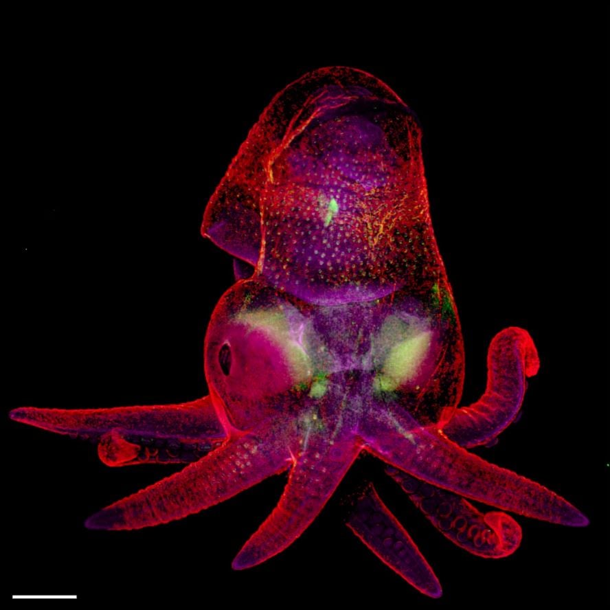
-
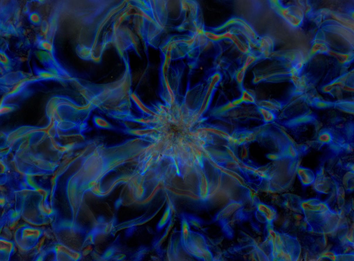
Best Technology: for Thomas Parton, a PhD student at the Department of Chemistry in the University of Cambridge.
His image "Colour from chaos: order and disorder" shows the intense colors seen in a micrograph created by cellulose nanocrystals, tiny slivers of carbohydrate extracted from cotton. Each individual nanocrystal is just about 200 nanometres long, which is too small to be seen under most conventional microscopes. However, under the right conditions these nanocrystals pack together into micron-scale screw-shaped arrangements that only reflect certain colors – in scientific terms, the color reflected has a wavelength corresponding to the pitch (periodicity) of the screw.Thomas studies ways to use cellulose as a more sustainable source of colorants, instead of the dyes and plastics we currently use. The "tendrils" seen in the image are created when the cellulose crystals try to assembly around micelles (clusters of molecules called surfactants, which are the main component of soaps and detergents). He uses these surfactants to move the cellulose crystals around and control their color. This image is something of a failure, he said, as he didn't achieve very uniform color, it looked very beautiful nonetheless! The image was taken on an inverted optical microscope in reflection mode with crossed polarizers. The image was formed by stitching together nine high-resolution images taken at 50x magnification.
-
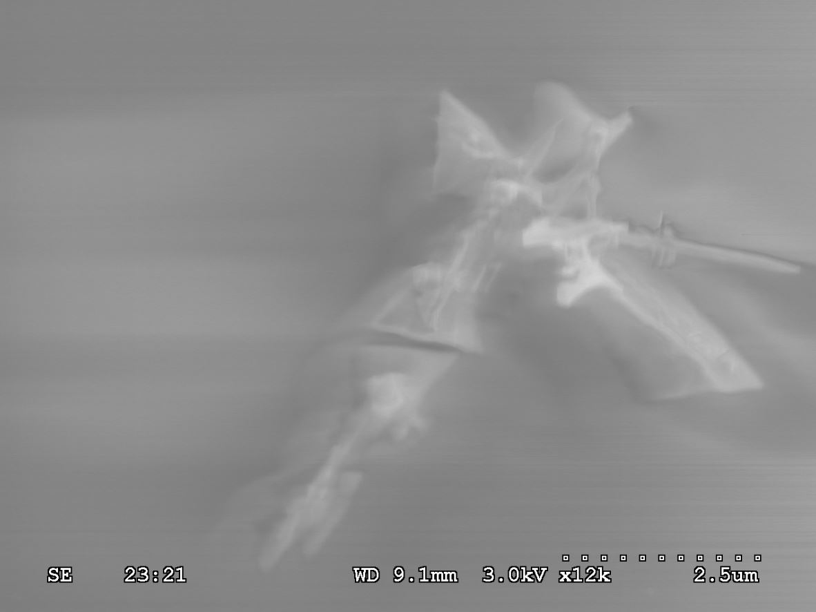
Best humoristic: for Xiaosheng Cai, a PhD student at the department of Material Sciences of the University of Oxford.
His image "The two-dimensional Jedi Knight" presents a two-dimensional nanosheet composed of tungsten disulphide, which is made by chemical vapor deposition. It looks like the Jedi Knight, wearing the hooded robe with the traditional weapon of the light-saber, in the Star Wars universe. The size is of 5 x 5µ captured by scanning electron microscopy. When the bulk tungsten disulphide downscales to one monolayer, it becomes a direct band gap semiconductor of 2.02eV with superior properties including valleytronic performance, photo-luminescence, and high carrier mobility, which is suitable for application in electronics and optoelectronics.
-
People's choice award: the public votes agreed with the jugdes' Best Scientific impact award, as Martyna Lukoseviciute with her "Hidden beauty of an octopus embryo" was voted as the winner.
-1.jpg)
-
People's technology award: the other prize chosen by attendees went for the same author as the Best Technology award Thomas Parton, for his image "Colour from chaos: order and disorder", who unfortunately couldn't make it to the event to collect his awards.
Special mentions
Considering the large number of eye-catching creative material submitted, the judges announced some entries that deserved a special mention. These were also presented during the award ceremony and were:
- Dr Stefania Kapsetaki, a postdoc from the Department of Zoology at the University of Oxford for "Exploring my universe"
- >Anna Valchanova, an undergraduate student from University College London for "Ebola Virus Glycoprotein Fragment" (pictured)
- Dr Zeynep Okray, a postdoc from the Department of Physiology, Anatomy and Genetics at the University of Oxford for his image "Achoo!"/li>
- A joint submission from Joint submission: PhD student Jiale Wang, Professor Susannah Speller and Dr. Sangeeta Santra (presented) from the Department of Materials at the University of Oxford for their image "Seasons"
-
Mayur Saxena, an MSc student from the Department of Computer Science at the University of Oxford for "Learning to love"
mod-1.jpg)
If you are keen on exploring the interface between Art and Science, keep an on the ONI website and twitter account for news. Alternatively, you can always get in touch with our team.